Before surgery photos of dogs with Dermoid Sinus
Unless otherwise noted, all photographers were taken by and remain the property of Elizabeth Akers.

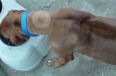
This is what the skin can look like, once the neck has been shaved and the neck skin has been “scruffed”. You will see the dimpled depression in the skin and the small hole that resembles a dark pore. This is the “exit” hole of the Dermoid.
A large lump caused by the infected tube of the DS - in this case there were two dermoids next to each other - Abby, the girl seen in this photo as well as several others on this site, is now living a happy and contented life in Washington.
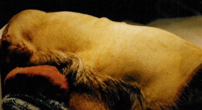
10 week old GSDxRR mix - DS lump visible, the tube was palpable at three days of age. Bliksem lived with me until his death in May, 2004. He was 10 years of age.
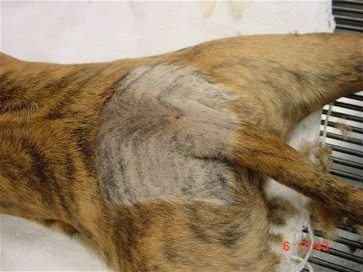
8 week old PitbullxRR mix - DS found in the tail area. This dog had several removed and later had his tail amputated due to the DS regrowth. Brodie lives in Marin county.
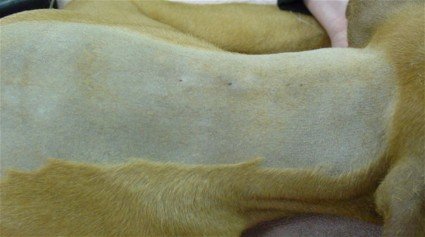
Two distinct DS “holes” which are actually the exits of the Dermoids - Shadow is living happily with her family in California.
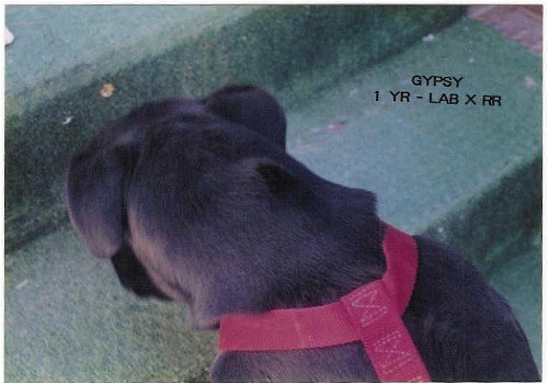
One year old LabxRR mix. The shelter vet told me that this lump was simply a vaccine reaction. The DS was successfully removed a few weeks later. Gypsy went to live with a family in LA.
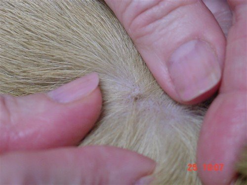
The surface hole may look like this prior to being shaved.
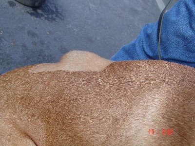
An infected Dermoid located mid back on a ridgeless mixed breed. He had a second Dermoid in the neck. His sister who is ridged, had a Dermoid in her neck.
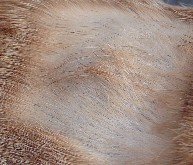
Infected DS prior to surgery
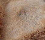
Infected DS prior to surgery
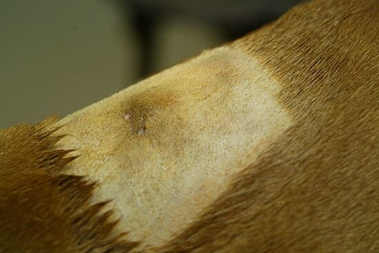
Shaved area showing the lump of an abscessed dermoid with the telltale “vampire” bite holes marking the dermoid.

Shaved area showing an abscessed DS on the head.
